|
|
|
|
|


Fetal, Pregnant Women and Infants Numerical Models

The models distributed in this project are licensed under a Creative Commons Attribution-Noncommercial-Share Alike 2.0 France License.
General Overview
The aim of the ACTE project is to develop reliable 3D models to be used for numerical dosimetry studies. These models were built using different approaches combining image segmentation, growth modeling, smooth meshes deformation and physical deformation. The broad ideas of the approaches developped during the course of this project are summarized next.Multi-organs segmentation
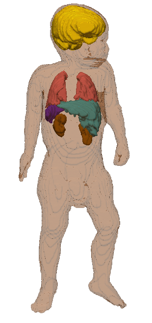
A shape-guided multi-region variational region growing framework for extracting simultaneously thoracic and abdominal organs on 3D infants whole body MRI has been developed. To enable the simultaneous segmentation of multiple organs, we introduce a segmentation framework on a graph of supervoxels that combines supervoxels intensity distribution weighted by gradient vector flow value and a shape prior per tissue. The intensity-based homogeneity criteria and the shape prior, encoded using Legendre moments, are added as energy terms in the functional to be optimized. The intensity-based energy is computed using both local (voxel value) and global (neighboring regions mean values, adjacent voxels values and distance to the neighboring regions) criteria. Inter-region conflict resolution is handled using a weighted Voronoi decomposition method, the weights being determined using tissues densities. The energy terms of the global energy equation are weighted using an information on growth direction and on gradient vector flow value.|
|
|
|
|
3D MRI image |
Automatic Segmentation |
3D reconstruction of the infant tissues |