|
|
|
|
|


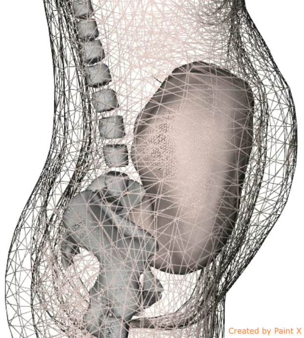
Fetal, Pregnant Women and Infants Numerical Models

The models distributed in this project are licensed under a Creative Commons Attribution-Noncommercial-Share Alike 2.0 France License.
General Overview
Several international scientific organizations and institutes, such as the World Health Organization (WHO) and the COST 281 have expressed the need for developing numerical models of the human body to enable studies of the interactions between radio-frequency electromagnetic waves and biological tissues. Several previous works have focused on developing models of adults and children heads, based on magnetic resonance imaging (MRI) data. New usage habits of mobile phones (hand free kits, ...) and the introduction of new technologies based on electromagnetic fields (Wifi, ...) have raised the need to develop whole body numerical models, based on medical images. With the advent of both obstetrical and whole body imaging, models of the fetus and the pregnant woman and of the infant are now considered.Description of both fetal projects are made on the Fetal Projects pages and the infant modeling project in presented on the Infant Projects page.